

You smile in a photo and notice it straight away: the gums above your front teeth are brown or almost black, not the pink you see in toothpaste ads. Maybe a friend has pointed it out, or you have started covering your mouth when you laugh. Then the questions arrive. Is something wrong with my gums? Is it from smoking, a filling, or something more serious?
The reassuring part first: in most people, darker gums are simply natural pigment. The same melanin that gives skin its color also sits in the gums, and many people, especially those with medium or darker skin, have gums that are naturally brown in places. This is not a disease and does not harm the teeth. A smaller group of dark areas comes from smoking, old metal fillings or crowns, or medicines, and a few spots need a closer look. Telling these apart is the first step.
This article helps you do that. You will find a one-minute self-check, a table of causes and how to tell them apart, the features that mean a spot should be checked, how smoking affects gum color, the options for evening out gum color and what research says about the color coming back, common myths, and what happens at a consultation.
In short: dark gums are usually natural pigment and harmless. An even band of color on both sides that you have had for a long time is rarely a concern. A single new spot, a spot that grows or changes, or one that is raised or sore should be checked by a dentist. If you simply want a more even gum color, laser depigmentation can remove the thin pigmented layer after the cause has been checked.
Is my dark gum color normal? A one-minute self-check
Stand at a mirror in good daylight, lift your upper lip and pull down your lower lip. Look at the color, the pattern and the edges. Then see which group below fits you most closely.
Usually natural pigment: no rush
- The color is a fairly even brown band across the gums of several front teeth, on both sides.
- You have had it for as long as you can remember, or it looks similar in old photos.
- The edge of the gum right next to the teeth is often lighter pink.
- Family members have similar gum color.
- The gums are firm, do not bleed easily and are not sore.
Worth mentioning at your next dental visit
- You smoke, and the darker color is mostly on the gums of the lower front teeth.
- There is a gray or blue-black spot next to an old silver filling or under a missing tooth area.
- There is a thin dark line along the gum edge of a crown.
- You started a new long-term medicine and later noticed color changes in the mouth or skin.
- The gums are also red, puffy or bleed when you brush.
Worth booking a check soon
- A single dark spot that appeared recently, or one you are not sure about.
- A spot that is getting bigger, darker, or changing shape or color.
- Uneven color within one spot, an irregular edge, or a raised or lumpy surface.
- A sore, ulcer or bleeding at the spot that has not healed after about 3 weeks.
- New dark patches in the mouth together with tiredness, weight loss or darker skin.
The last group is not a reason to panic. Most spots that are checked turn out to be harmless. A check simply gives you a clear answer, which is more useful than worrying.
Why some gums are darker than others
Gum color comes from a mix of things: how thick the surface layer is, how many small blood vessels sit underneath, and how much melanin the pigment cells make. Melanin is made by cells called melanocytes in the lowest part of the surface layer. Everyone has roughly similar numbers of these cells. What differs is how active they are, which is why skin tone and gum tone often go together.
Research on natural gum pigment shows a typical pattern. A study of gum pigment in people with darker skin found that the most common form was a broad, continuous ribbon of color across the attached gums from the front teeth to the canines, symmetrical on both sides, with the gum edge next to the teeth still pink. Age also made a difference. If your gums look like this, it matches natural pigment rather than disease.
Pigment can also increase for other reasons. Tobacco smoke stimulates the pigment cells. Some medicines and some hormone conditions do the same. Metal particles from old fillings can sit in the gum like a small tattoo. Bleeding under the surface can look dark for a while, like a bruise. Each of these looks a little different, which is why the pattern and history matter so much. For example, the NHS lists areas of skin that are darker than the surrounding skin among the symptoms of Addison disease, a hormone condition, alongside tiredness and weight loss.
How common are the less usual causes? In a large review of tissue samples from two oral pathology services, pigmented lesions made up about 1 in 100 of all mouth lesions that were sampled. Among those, the most common were gray marks left by particles of old silver filling material, then flat freckle-like spots and natural pigment. Melanoma was about 2 in 100 of the pigmented lesions. In other words, it is rare, but it is the reason a new or changing spot deserves a look.
Causes of dark gums and how to tell them apart
Use this table together with the self-check. A dentist looks at the same features, plus your history and, when needed, an X-ray or a small tissue sample.
| Cause | How to tell | What to do |
|---|---|---|
| Natural pigment (physiologic pigmentation) | Even brown band on both sides, present for a long time, often similar in family members. | No treatment needed for health. Depigmentation is an option if the color bothers you. |
| Smoking (smoker melanosis) | Brown patches mostly on the gums of the lower front teeth, more with heavier smoking. | Stopping smoking is the main step. Tell your dentist, especially if a patch keeps darkening. |
| Secondhand smoke | Similar patches in children or adults who do not smoke but live with smokers. | A smoke-less home helps. Mention it at the child dental check. |
| Gray mark from an old silver filling | Flat gray, blue or black spot next to an old silver filling, crown or a past extraction site. Does not change. | Harmless once confirmed. Have it checked once, because it can look like other spots. |
| Dark line at an old crown | Thin gray line where a metal-based crown meets the gum, more visible if the gum has pulled back. | Ask whether replacing the crown with one that has no metal suits you. |
| Flat freckle-like spot (melanotic macule) | Small, flat, evenly brown spot with a clear edge, often on the lip or gums. | Usually harmless. Have a new spot checked once and watch for change. |
| Some medicines | Gray, blue or brown areas that appear after starting a long-term medicine. | Do not stop the medicine yourself. Tell your doctor and dentist the name of the medicine. |
| Hormone and body conditions | New patches in the mouth together with darker skin, tiredness or weight loss. | See a doctor for a general check. Your dentist can note what is in the mouth. |
| Bruise or blood under the gum | Dark red to purple area after an injury or hard brushing. Fades over days to weeks. | Brush gently. If bruises appear without a reason, see a doctor. |
| Inflamed gums | Dark red or purplish, swollen gum edges that bleed when you brush. | Clean gently and thoroughly. Book a gum check if it does not settle. |
| Pigmented spot that needs a biopsy | New, growing, uneven in color, irregular edge, raised, or ulcerated. | Have it examined soon. A small tissue sample gives a clear answer. |
See how a gum examination works
Harmless pigment or a spot to check?
Dentists use a simple set of questions to decide whether a dark area can be watched or should be examined more closely. You can use the same questions at home. They are not a diagnosis, but they help you decide how soon to book.
| What you see | Usually harmless when | Have it checked when |
|---|---|---|
| How many areas | Many areas, spread evenly on both sides | One area on its own |
| How long it has been there | As long as you remember | New, or you are not sure |
| Change | Looks the same in old photos | Growing, darker or changing shape |
| Color | Even light to dark brown | Several shades in one spot, or very black |
| Edge and surface | Flat, smooth, soft edge | Irregular edge, raised, lumpy |
| Symptoms | No symptoms | Sore, bleeding or an ulcer that has not healed in about 3 weeks |
The NHS advises seeing a dentist or doctor for a mouth ulcer that lasts more than 3 weeks, a red or white patch, or a lump in the mouth. Early checks make any treatment simpler, and most of these signs turn out to have a harmless cause.
Smoking and dark gums
Smoking is the most common cause of extra gum pigment that you can change. In a classic Swedish study, about a quarter to a third of smokers had this smoker melanosis, and the patches were almost always on the gums of the lower jaw, especially in front of the canines and incisors. People who had it smoked more than smokers who did not.
Because smoker melanosis usually starts in adult life and can slowly darken, dental references note that it should be told apart from other pigmented spots, which is one more reason to show it to a dentist once. Smoke also affects people around you. A study of non-smoking children and young adults with smoking parents found more gum pigment and nicotine by-products in their urine.
If you want lighter gums, start here
- Decide on a quit date and tell the people around you.
- Call Thailand national quitline 1600 for telephone advice, or ask your doctor about medicines that help.
- Make your home and car smoke-less, especially if children live with you.
- Keep brushing and cleaning between your teeth every day. Quitting can make gums bleed a little more at first as blood flow returns.
- If you plan depigmentation, it makes sense to quit first, because ongoing smoking is the cause that keeps making new pigment.
Can gum color be evened out? The options
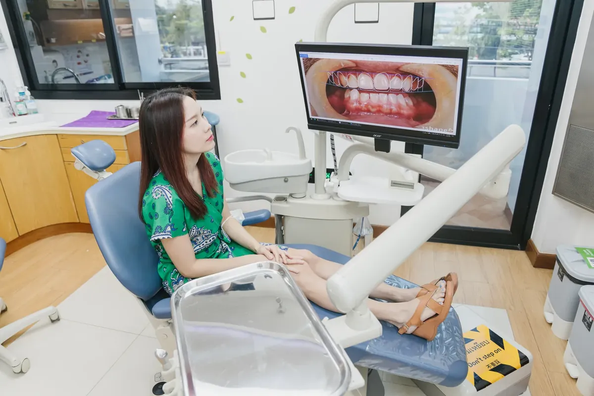
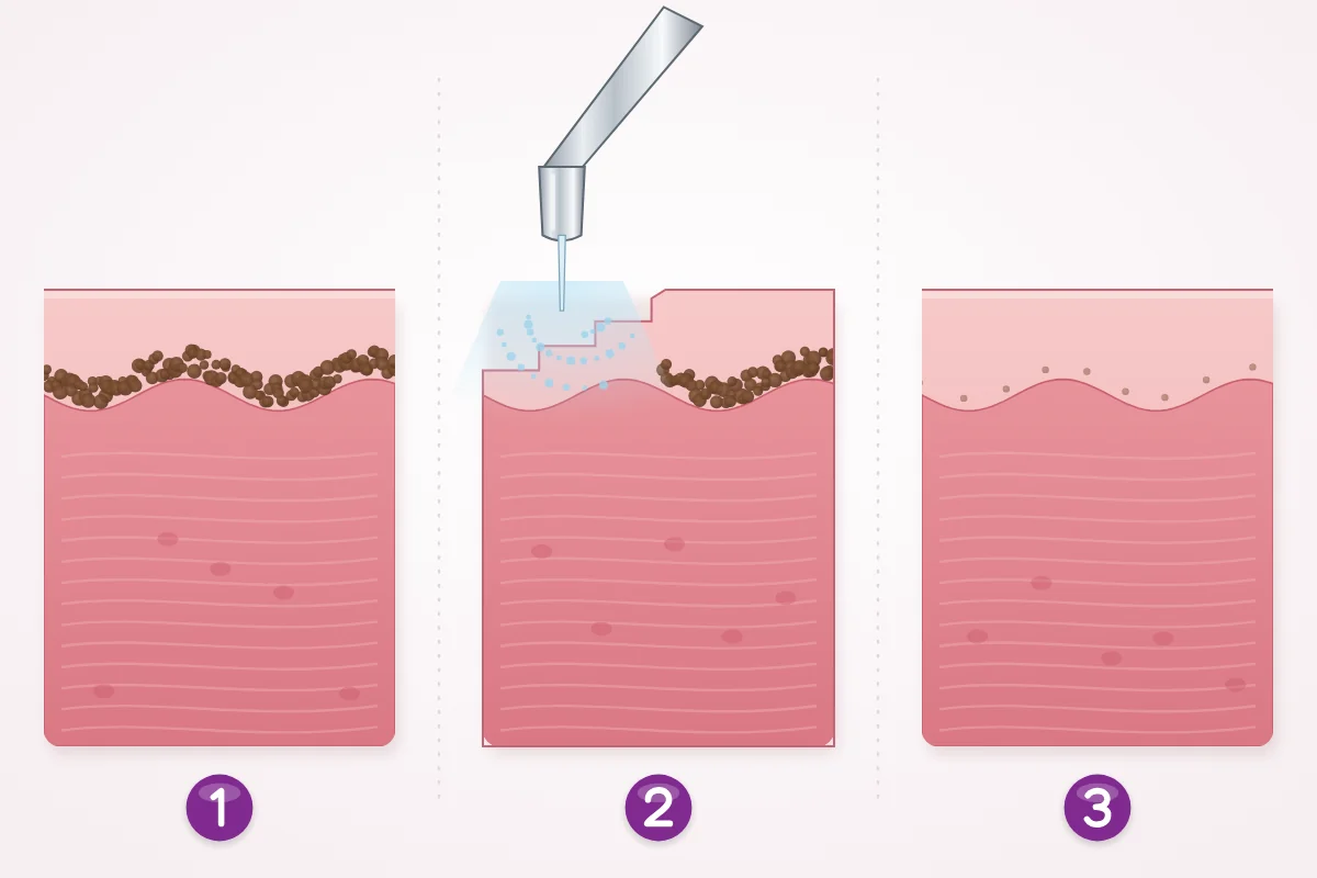
Yes, when the dark color is natural pigment or smoker pigment and you would like a more even color. The treatment is called gum depigmentation. Pigment sits only in the thin surface layer of the gum, so the idea is to remove that layer carefully. As the surface heals, new gum tissue grows in with less pigment, and the color usually looks more even.
Depigmentation is a cosmetic choice, not a medical need. It is not suitable for spots whose cause is still unclear, which should be examined first, or for gray marks from old silver fillings, where the color sits deeper. Gums that are inflamed are treated first, so the result is easier to judge.
| Method | How it works | What research says |
|---|---|---|
| Laser | Light energy removes the pigmented layer in very thin steps. | The most studied method. Less pain after treatment than a scalpel in pooled trials. |
| Scalpel (surgical stripping) | The surface layer is peeled away by hand. | The long-standing standard. Color results similar to laser. |
| Bur abrasion | A rotating instrument smooths away the surface. | Highest recurrence in one large review. |
| Electrosurgery or freezing | Heat or cold destroys the pigmented layer. | Low recurrence in case reports, but harder to control depth. |
| Chemical agents | A chemical is applied to peel the surface. | Rarely used now because it is hard to control. |
Two reviews help set expectations. A large review of 61 publications, mostly case reports, found that the color came back in a small share of cases with laser, electrosurgery or freezing, and most often after bur abrasion. A later review and meta-analysis of 25 clinical trials found laser and scalpel gave similar color results and similar recurrence overall, with less pain after laser. It also noted some return of color by 6 months in some laser studies. The honest summary: the color usually looks much more even, and some pigment can return over time, especially if the cause, such as smoking, continues.
At Kitcha Dental Clinic, gum depigmentation is done with a Waterlase dental laser, which works together with a fine water spray. The laser removes only the thin pigmented surface layer, and the water spray helps keep the area cool. Your dentist first checks the cause of the dark areas and explains what result to expect before you decide.
Read about laser treatment at the clinic
A step-by-step plan for dark gums
Whether you want to treat the color or simply want peace of mind, these steps keep things in a sensible order.
| Step | What to do | Why it helps |
|---|---|---|
| 1. Photograph | Take a clear photo of your gums in daylight. Repeat in about a month. | Shows whether anything is changing, which is the most useful clue. |
| 2. List the history | Write down smoking, medicines, old fillings or crowns near the area, and when you first noticed it. | Most causes can be identified from history and appearance. |
| 3. Get it checked | Show single, new or changing spots to a dentist first. | Rules out the uncommon causes before any cosmetic step. |
| 4. Treat the gums | Brush gently at the gum line and clean between teeth every day. | Healthy gums heal better and make the result easier to judge. |
| 5. Remove the cause | Stop smoking, or discuss an old crown margin with your dentist. | Lowers the chance that the color comes back. |
| 6. Decide on depigmentation | Ask what result to expect, how the area is cared for afterward and how fees work. | You decide with clear information. |
Looking after the gums after depigmentation
The treated area may feel tight or a little tender for a short time, depending on how large the area is. Your dentist gives you aftercare instructions that match your treatment and tells you when to come back. General advice after gum procedures includes the following.
- Follow the brushing instructions for the treated area exactly, and keep brushing the rest of your teeth as usual.
- Choose soft, lukewarm food at first and avoid very spicy, very hot or crunchy food on that side.
- Avoid smoking, which slows healing and brings pigment back.
- Contact the clinic if pain increases after the first days, or there is swelling or a fever.
The number of visits and how long the gums take to settle depend on the size of the area and your gums, so your dentist tells you for your case.
Common myths about dark gums
| Myth | Fact |
|---|---|
| Dark gums mean the gums are unhealthy. | Natural pigment says nothing about gum health. Healthy gums are firm and do not bleed, whatever their color. |
| Whitening toothpaste or gum creams can lighten dark gums. | Pigment sits inside the gum tissue. Toothpaste works on tooth surfaces and does not reach it. |
| Rubbing lemon, salt or bleach on the gums helps. | These can burn or wear the gums and cause gum recession. Avoid them. |
| Every dark spot is a sign of cancer. | Most dark spots are harmless. A new or changing spot simply needs a check to be sure. |
| Once removed, the color can never come back. | Research shows some pigment can return over time, more often if smoking continues. |
| Smokers have pink gums, so smoking does not affect gum color. | Not everyone reacts the same way, but studies link smoking clearly with extra gum pigment. |
| A gray mark from an old filling is harmful and must be cut out. | Once confirmed, it is considered harmless and usually needs no treatment. |
When to see a dentist or doctor soon
These signs do not mean something serious is happening. They mean a professional look is the sensible next step.
- A dark spot that is new, growing, getting darker or changing shape.
- A spot with several colors, an irregular edge, or a raised surface.
- An ulcer, lump, or a red or white patch that has not healed after about 3 weeks.
- Dark patches in the mouth together with darker skin, tiredness, dizziness or weight loss: see a doctor for a general check.
- Bruise-like patches in the mouth that appear without injury, especially with nosebleeds or bleeding gums: see a doctor promptly.
Need to be seen soon? Contact the clinic
What happens at a consultation, step by step

- Talk it through. Your dentist asks when you noticed the color, whether it has changed, and about smoking, medicines and past dental work.
- Examine the gums. Your dentist looks at the color, pattern and surface of the dark areas, and checks the gums themselves, measuring the gum pockets when needed.
- Find the cause. Natural pigment and smoker pigment are usually clear from the pattern. A spot next to old fillings or crowns may be checked with an X-ray. A spot that is unusual is examined further.
- Explain the options. If the color is suitable for depigmentation, your dentist explains how the Waterlase laser is used, what result to expect, aftercare, and the fee before you decide.
- Treat in the order that makes sense. Inflamed gums are treated first. If an old crown edge is the cause, your dentist discusses whether a crown with no metal, such as zirconia or all-ceramic, suits you.
- Depigmentation and follow-up. The laser removes the thin pigmented layer. Your dentist gives aftercare instructions and arranges follow-up as needed. The number of visits and healing time are explained for your case.
Gum care at the clinic is planned by dentists with further training in periodontics, including a master degree in periodontics and a doctorate. Most laser work is an added fee on top of the main treatment, and some items have their own price per tooth. Your dentist confirms the fee before treatment.
If this sounds like you: what to do
These are example situations, not patient stories, to help you find the next step that fits.
- Your gums have always been brown in a band above the front teeth and you dislike it in photos: this matches natural pigment. If the color bothers you, a consultation about laser depigmentation is a reasonable next step.
- You smoke and the gums of your lower front teeth have darkened: stopping smoking is the first step. Show the area to a dentist once, and decide about depigmentation after you have quit.
- There is a flat gray spot next to an old silver filling: it may be a mark from the filling material. A dentist can confirm it, and if it is, it usually needs no treatment.
- A gray line shows at the edge of an old crown on a front tooth: ask whether a crown with no metal suits you, especially if the gum has pulled back.
- You noticed one new black spot that seems larger than last month: book a check soon. A clear answer brings peace of mind.
- Your child has brown patches on the gums and someone at home smokes: make the home smoke-less and mention it at the next dental check.
- You plan veneers or crowns and want the gums to match: mention the gum color at the planning stage, so the order of treatment can be planned together.
Frequently asked questions about dark gums
Are dark gums dangerous?
Usually not. Most dark gums are natural pigment or pigment from smoking. The ones to have checked are single spots that are new, growing, changing, raised or sore.
Can dark gums go back to pink on their own?
Natural pigment does not fade on its own, because it is part of how your body makes color. Pigment linked to smoking is caused by the smoke, so stopping removes the trigger. A bruise fades within days to weeks.
Does laser depigmentation hurt?
The area is numbed with local anesthetic when needed, so you feel little during treatment. Afterward it may feel tight or tender for a short time. In pooled trials, people had less pain after laser than after scalpel removal.
Will the dark color come back?
It can, partly and slowly. Research shows recurrence varies between people and methods, and is more likely if the cause, such as smoking, continues. Many people find the result stays much more even than before.
Can I lighten my gums at home?
Not safely. Pigment sits inside the gum, out of reach of toothpaste, gels or home remedies. Rubbing acids or bleach on the gums can damage them. What you can do at home is stop smoking and keep the gums healthy.
Why are my gums dark around a crown?
Often the metal layer inside an older crown shows through thin gum, or the gum edge has pulled back and shows a dark line. Sometimes the gum is inflamed. A dentist can tell which it is and whether a crown with no metal is a suitable option.
My child has dark gums. Should I worry?
Many children have natural gum pigment, especially if parents do. A bluish swelling over a tooth about to come through is usually a small blood-filled bump that settles once the tooth erupts. Secondhand smoke is linked to gum pigment in children. Mention any concern at the next dental check.
Can everyone have gum depigmentation?
Not always. Spots with an unclear cause are examined first, inflamed gums are treated first, and thin gums or certain health conditions may change the plan. Your dentist explains whether it suits you and what result to expect.
Summary
Dark gums are usually natural pigment, are not a disease and do not harm your teeth. The aim is to tell harmless pigment apart from the few spots that need a check.
- An even, long-standing band of color on both sides is rarely a concern.
- Have a single new, growing, changing, raised or sore spot checked soon.
- Smoking is a common cause you can change, and it also affects children at home.
- Laser depigmentation can make gum color more even after the cause is checked. Some color may return over time.
- Skip home bleaching and acids on the gums. Healthy gums and a smoke-less life help most.
References
Ko E, Panchal N. Pigmented lesions of the oral cavity. Dermatol Clin, 2020
Brown FH, Houston GD. Smokers melanosis: a case report. J Periodontol, 1991
NHS (UK): Mouth cancer, symptoms