Your dentist says you need a 3D scan before an implant, or before a wisdom tooth comes out. Straight away the questions start: is that a lot of radiation, is it the same as a hospital CT, did the small X-ray not show enough, and is it really necessary?
The reassuring part first: a dental CBCT (cone beam CT) scan uses more radiation than a regular dental X-ray, but generally less than a medical CT scan. Dental and radiation safety bodies agree that when a scan answers a real clinical question, its benefit outweighs the small radiation risk. That is why dentists use it only when a flat X-ray cannot give the information needed.
This article explains the different dental X-rays and what each one shows, when a 3D scan helps and when it does not, radiation doses in plain numbers, what to know if you are pregnant or the scan is for a child, what the scan is actually like, and what happens at our clinic, including booking a CBCT scan on its own.
In short: a CBCT scan is a 3D X-ray of the teeth and jaws. It shows bone, roots, nerves and the sinus in depth, which flat X-rays cannot. It is used when a treatment decision depends on that detail, such as implant planning or a wisdom tooth near the nerve. The dose is higher than a regular dental X-ray and generally lower than a medical CT, so it is taken only when needed.
3D scan or a regular X-ray? A quick self-check
Only your dentist can decide which image you need, but this list helps you understand the reason and ask good questions.
A regular X-ray is usually enough
- A routine check-up, or looking for decay between the teeth.
- A filling, or checking an old filling or crown.
- Most first-time root canal treatment on a tooth with simple roots.
- Checking the gums and bone level around the teeth.
- An overview of all the teeth, including wisdom teeth that sit away from the nerve.
Your dentist may suggest a 3D scan
- You are planning a dental implant, and the bone height, width and nearby nerve or sinus need to be measured.
- A lower wisdom tooth looks close to the nerve on a panoramic X-ray.
- A tooth has curved or extra canals, a suspected crack, or needs root canal retreatment.
- Surgery near the root tips, nerves or sinus, such as root-end surgery.
- An impacted tooth, such as a canine, whose exact position matters for treatment.
Tell your dentist first
- You are pregnant, or might be.
- The scan is for a child or teenager.
- You had a dental CBCT or X-ray recently, at any clinic. Bring or send it, because it may be usable.
- You find it hard to keep still for a short time, or you feel anxious in enclosed or noisy machines.
Types of dental X-rays and what each one shows
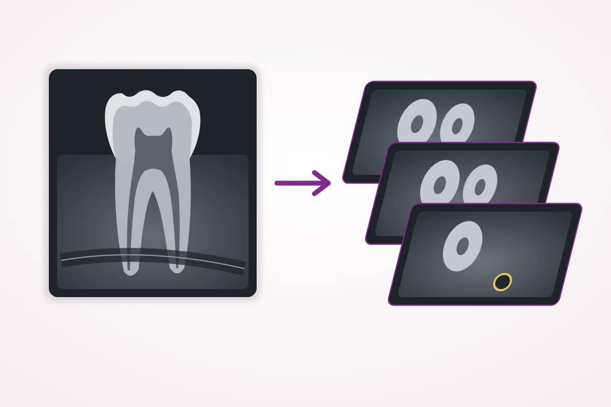
Dental X-rays fall into two groups. Intraoral X-rays use a small sensor placed inside the mouth and show a few teeth in fine detail. Extraoral X-rays are taken with the sensor outside the mouth and show a wider area, such as all the teeth and both jaws. Dense tissue such as teeth and bone absorbs more X-rays and looks white, while softer tissue looks darker.
| Type of X-ray | What it shows | Typical use |
|---|---|---|
| Bitewing (small film inside the mouth) | The crowns of upper and lower back teeth together; usually not the roots | Decay between teeth, under old fillings, and bone level near the gum line |
| Periapical (small film inside the mouth) | One or two whole teeth, from crown to root tip, and the bone around them | Root canal treatment, infection at the root tip, bone loss around a tooth |
| Panoramic (OPG, outside the mouth) | All the teeth, both jaws, the jaw joints and the sinus in one flat image | Overview, wisdom teeth, planning braces or dentures, first assessment for implants |
| Cephalometric (side view of the head) | The skull, both jaws and the teeth seen from the side | Planning orthodontic treatment and jaw surgery |
| CBCT (3D cone beam CT) | Teeth, bone, roots, nerve canals and the sinus in three dimensions, in slices from any direction | Implant planning, wisdom teeth near the nerve, complex root canals, surgery planning |
Flat X-rays squeeze everything into one image, so structures in front and behind overlap. A panoramic X-ray can show that a wisdom tooth root and the nerve canal cross, but not whether the nerve runs in front of, behind or right against the root. CBCT answers that by showing depth.
What CBCT is, and how it differs from a hospital CT
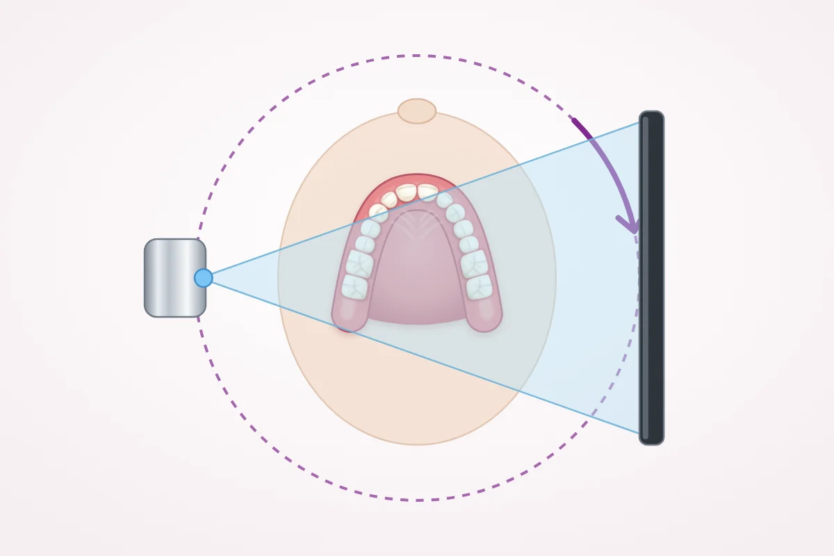
CBCT stands for cone beam computed tomography. The machine sends a cone-shaped X-ray beam through the area being examined while it turns once around your head. It records hundreds of flat images on the way round, and a computer combines them into a 3D picture. Your dentist can then look at the teeth and jaws in slices from top to bottom, side to side and front to back.
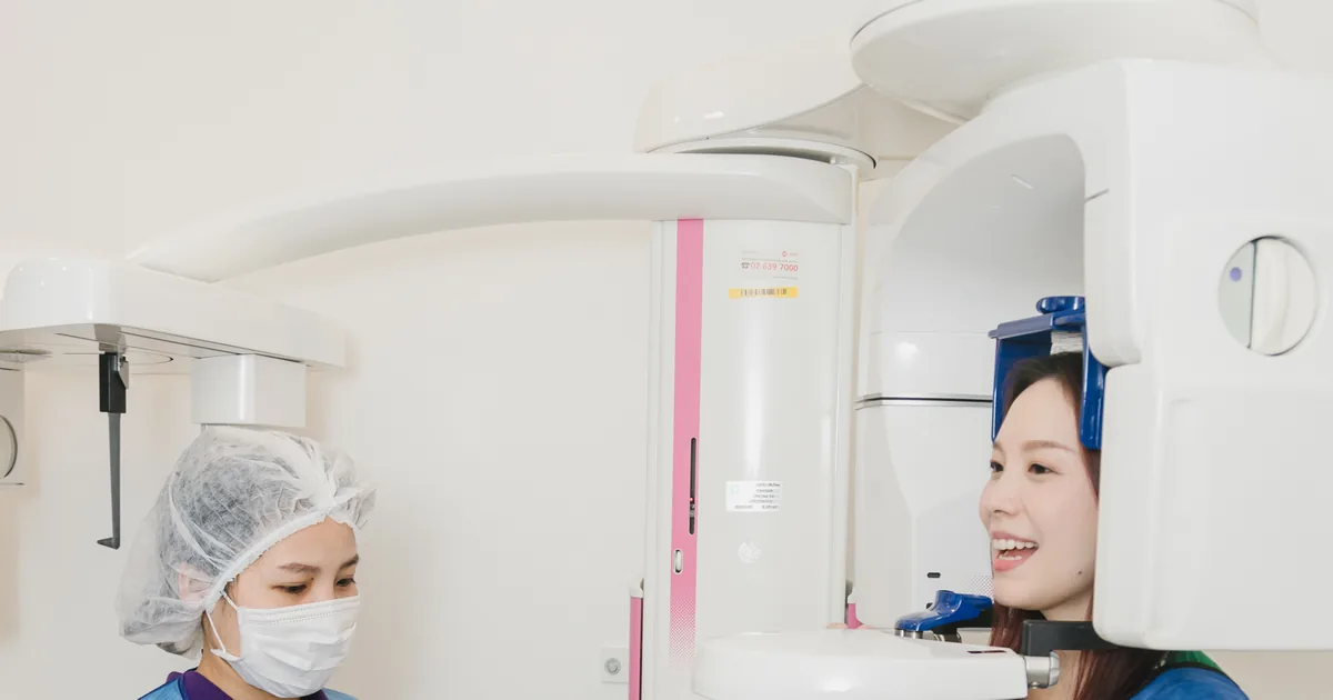
Unlike a hospital CT, a dental CBCT machine is small enough for a dental clinic, and you sit or stand rather than lie inside a tunnel. The scan area can be set from one or two teeth up to both jaws, and a smaller area generally means a lower dose.
| Feature | Dental CBCT | Medical CT |
|---|---|---|
| Position | Sitting or standing; the machine turns around your head | Lying on a table that moves through a ring |
| Radiation dose | Higher than regular dental X-rays, generally lower than medical CT | Generally higher |
| Bone and teeth detail | Very good for teeth, roots and jaw bone | Good for bone, and much better for soft tissue |
| Soft tissue such as muscles and glands | Limited | Shown in detail |
| Where it is done | Dental clinics and hospitals | Hospitals and imaging centers |
When dentists use a CBCT scan
Dental and radiology guidance says CBCT should be used when it gives information that lower-dose X-rays cannot, and when that information changes the plan. These are the situations where it is most often useful.
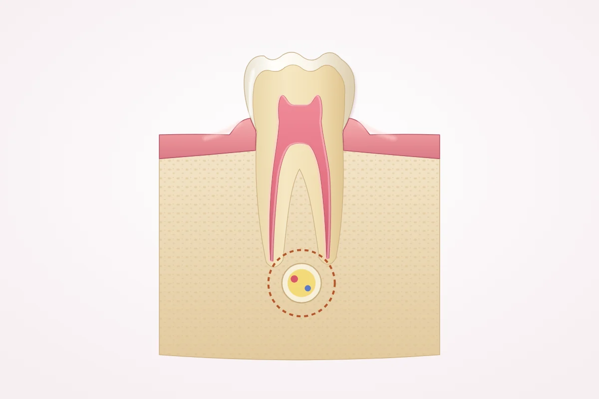
| Situation | What the 3D image adds |
|---|---|
| Dental implants | Measures bone height and width, shows the nerve canal and sinus floor, and helps choose implant size and position. A radiology academy recommends cross-sectional imaging for implant sites, with CBCT as the method of choice. |
| Bone grafting and sinus lift | Shows how much bone is missing and where, so the dentist can tell whether grafting is needed |
| Wisdom teeth close to the nerve | Shows exactly where the nerve runs in relation to the roots. In a review of studies, CBCT predicted nerve exposure during surgery more accurately than a panoramic X-ray. |
| Complex root canal treatment and retreatment | Helps find extra or curved canals, cracks and the extent of infection around the root |
| Impacted or extra teeth | Locates the tooth and its distance from neighboring roots |
| Jaw, sinus and jaw joint questions | Shows the bony parts of the jaw joint, cysts in the jaw and the sinus floor |
| Dental injuries | Helps show root fractures and damage to the bone around teeth |
For a routine check-up, a filling or most simple root canals, a CBCT scan usually adds little, and flat X-rays are the right choice. A scan is not a general screening test.
How much radiation is a dental CBCT scan?
Everyone receives natural background radiation every day, from the ground, building materials and space. In the United States the average is about 3 millisieverts (mSv) a year, a little under 0.01 mSv a day. Comparing a scan with that daily background is an easy way to picture the dose.
| Exam | Typical effective dose | About the same as natural background |
|---|---|---|
| Small dental X-ray inside the mouth | About 0.005 mSv | About 1 day |
| Panoramic X-ray | About 0.025 mSv | About 3 days |
| Dental CBCT, small area (adult average) | About 0.08 mSv | About 10 days |
| Dental CBCT, large area (adult average) | About 0.21 mSv | About 3 to 4 weeks |
| Chest X-ray | About 0.1 mSv | About 10 days |
| Medical CT of the head | About 1.6 mSv | About 7 months |
The CBCT figures are averages from a large analysis of published measurements. The same study found very wide ranges between machines and settings, so your own dose depends on the machine, the size of the area scanned and the settings chosen. The other figures are typical adult values published for patients by radiology societies.
How dentists keep the dose low
- Justify every scan: an image is taken only when it answers a question that affects your care.
- Choose the lowest-dose image that gives the answer, and use CBCT only when flat X-rays are not enough.
- Keep the scan area as small as the question allows. This is often called the ALARA principle: as low as reasonably achievable.
- Reuse recent images from earlier visits or other clinics whenever they still answer the question.
- Use settings suited to the patient size, especially for children.
Lead aprons and thyroid collars
Many people expect a lead apron. Updated recommendations from the American Dental Association in 2024 found that lead aprons and thyroid collars are no longer needed for dental X-rays, for patients of any age or health status, including pregnancy. Modern equipment with a tightly limited beam does the protecting, and a shield can block the beam and lead to a retake. Rules still differ between countries and clinics, so some places continue to use them.
Radiation from CBCT and X-rays inside the mouth at our clinic
Many patients feel worried about radiation before a dental X-ray, and it is always fine to ask. At our clinic, three things work together to keep the dose low: a CBCT scanner with an Ultra Low Dose mode, digital image receptors for X-rays inside the mouth, and taking images only when they are needed.
Our Planmeca ProMax CBCT has an Ultra Low Dose mode
The clinic CBCT scanner is a Planmeca ProMax, which takes both 3D CBCT scans and panoramic X-rays in one machine. It has the Planmeca Ultra Low Dose (ULD) mode which, according to the manufacturer, lowers the dose by reducing the tube current and shortening the X-ray pulse for each image, rather than by taking fewer images. Planmeca reports an average dose reduction of 77% compared with standard protocols, citing a study by Ludlow and Koivisto.
An independent study published in Dentomaxillofacial Radiology in 2023 measured a ProMax 3D Mid on a head phantom with a small scan area of 4 × 5 cm. The effective dose was 2.8 to 5.8 µSv (about 0.003 to 0.006 mSv) in Ultra Low Dose mode, compared with 16.0 to 30.9 µSv (about 0.016 to 0.031 mSv) in the standard mode, roughly six times lower. The authors concluded that low-dose protocols may be adopted routinely for diagnosis and treatment planning. These are study results on a phantom, not a measurement for each patient at our clinic.
Your dentist chooses the mode and the scan area for each case. Ultra Low Dose suits many planning and follow-up scans, while some tasks, such as looking at very fine detail around a root, need the sharper image of a standard setting. Either way, the scan area is kept as small as the question allows. For example, if only one tooth needs checking, the whole jaw does not need to be scanned.
The actual dose of each scan depends on the mode, the scan area and the patient size, so no single figure applies to everyone. The figures in the table above are averages from research covering many machines and are there for comparison. If you would like to know how large an area your scan will cover, ask your dentist before the scan.
Digital X-rays inside the mouth: phosphor plates and sensors
Small X-rays inside the mouth, such as bitewings and periapical X-rays, are taken at the clinic with digital receptors instead of film. There are two kinds: phosphor plates, which are thin and flexible like film, and sensors, which send the image straight to the computer. Guidance from the American Dental Association with the US Food and Drug Administration (2012) states that digital imaging provides an opportunity to reduce the radiation dose by a further 40 to 60 percent compared with film.
The image appears on a computer screen with no chemical film processing, so your dentist can enlarge it and explain it to you. How much dose is actually saved depends on suitable machine settings and getting a usable image the first time, which is why the team positions you and the receptor carefully before each exposure.
Only the images you need
- An image is taken when it answers a question that affects your treatment, not as a routine without a reason.
- If you have recent images from elsewhere, send or bring them, and your dentist checks first whether they can be used.
- If you are pregnant, tell your dentist that you are pregnant, and your dentist plans your care for you.
- You can ask anything before an image is taken, such as why it is needed and how large an area it covers.
In short: dental X-rays use a low dose of radiation, and the clinic combines three things to get the information needed with as little radiation as reasonably achievable: a CBCT scanner with an Ultra Low Dose mode that the dentist chooses when it suits the case, digital receptors, and taking images only when needed.
Pregnancy and children: can they have dental X-rays?
If you are pregnant
According to the American College of Obstetricians and Gynecologists, as reported by the American Dental Association, dental X-rays are safe during pregnancy. The doses are very low and the beam is aimed at the mouth, away from the abdomen. Many dentists still postpone X-rays that are not needed yet, and hospitals give special consideration to CBCT scans in pregnancy.
At our clinic: tell your dentist that you are pregnant, and your dentist plans your care for you.
If the scan is for a child
Children are more sensitive to radiation than adults and have more years ahead in which effects could appear. For that reason, guidance asks that CBCT be used for children only when it is essential, with settings adjusted to their size. Children and teenagers may still need regular dental X-rays more often than adults, because their teeth and jaws are changing. Parents can reasonably ask why the scan is needed, whether a lower-dose image would answer the question, and keep a record of the child imaging history.
A scan also needs the patient to keep still. A very young child who cannot stay still for a short time may not be suited to it.
What the scan is like, step by step
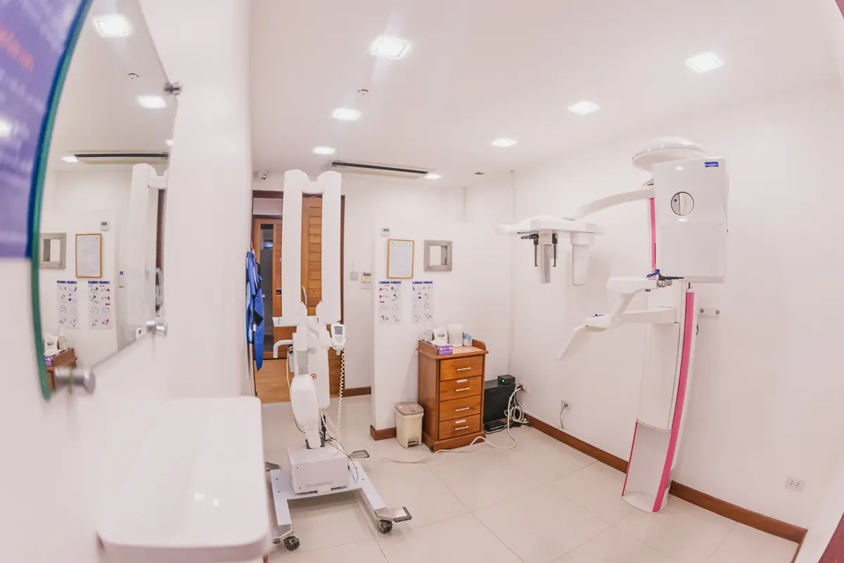
There is no special preparation. You do not need to stop eating or drinking, and you can take your usual medicines.
- Before the scan, tell the team if you are pregnant or might be, and mention any recent dental scans.
- Remove metal around the head and neck: earrings, necklaces, hairpins, glasses and hearing aids. Removable dentures and retainers usually come out too, so bring their case.
- You sit or stand in the machine. The team positions your head with a chin rest and sometimes a soft head support, and you may gently bite on a small guide.
- Keep very still and breathe normally while the arm turns once around your head. Nothing touches the inside of your mouth during the scan.
- The exposure itself is short: published patient information gives under 10 seconds for a small area, about 20 to 40 seconds for a full scan, and about 90 seconds once positioned on some machines.
- The computer builds the 3D image after the scan, which takes a little longer. You can go back to normal activities straight away.
- Your dentist reviews the images, explains what they show, and uses them in your treatment plan.
If you feel anxious in machines that move around you, say so. The team can explain each step before it happens, and you can practice staying still before the exposure starts.
What a 3D scan can and cannot show
CBCT is excellent for hard tissue: teeth, roots and bone. It is weaker for soft tissue such as muscles, glands and the gums themselves, which a medical CT or MRI shows far better. That is why it is used to answer questions about teeth and bone, not to examine every structure of the head.
- Metal such as crowns, posts, implants and some fillings can cause bright streaks and shadows called artifacts, which can hide detail next to them.
- Movement during the scan blurs the image, which is why staying still matters.
- Small cavities between teeth are often seen just as well, or better, on bitewing X-rays.
- A scan shows the current state only. Your dentist still examines your mouth, because some things can only be checked in person.
The large scan area also shows structures outside the teeth, such as the sinus and airway. Whoever reads the scan should look at everything in the image, not only the tooth in question.
Common myths about dental X-rays and CBCT
| Myth | Fact |
|---|---|
| A dental CBCT is the same as a hospital CT. | Both make 3D images, but dental CBCT is designed for teeth and jaws, is taken sitting or standing, and generally uses a lower dose. |
| A 3D scan is always better, so everyone should have one. | It is better only when the question needs depth. For check-ups and most fillings, flat X-rays answer the question with less radiation. |
| Dental X-rays are dangerous during pregnancy. | Dental X-rays are considered safe in pregnancy by obstetric and dental bodies. Tell your dentist, who decides what is needed now and what can wait. |
| Without a lead apron the X-ray is unsafe. | Current American Dental Association guidance says aprons and thyroid collars are no longer needed, because modern machines limit the beam. Local rules may still require them. |
| A CBCT scan can see everything, including gum disease and soft tissue problems. | It shows bone loss from gum disease, but not the gums themselves in detail. Gum health is checked by measuring the gum pockets. |
| If I had a scan at another clinic, I need a new one. | Not necessarily. A recent scan of the right area is often usable, and guidance encourages reusing earlier images. Your dentist decides after seeing it. |
Questions worth asking before a scan
Radiation safety agencies encourage patients and parents to ask a few simple questions. A good dental team is glad to answer them.
- What question will this scan answer, and how will the answer change my treatment?
- Would a lower-dose X-ray give the same information?
- How large an area will be scanned?
- Can my earlier X-rays or scan from another clinic be used instead?
- For a child: are the settings adjusted for a child size?
- Can I have a copy of the images for my records or another dentist?
CBCT at our clinic, step by step
A CBCT scanner is part of how treatment is planned at Kitcha Dental Clinic. Your dentist recommends a scan only when the plan needs it.
- Dental implants are planned with a 3D CBCT scan. Your dentist checks the bone, recommends an implant system and explains your timeline after the scan, and computer-guided surgery with a guide made from your scan is available when your dentist recommends it.
- Whether bone grafting or a sinus lift is needed is decided from the CBCT scan, and so is whether an implant can be placed right after an extraction.
- Wisdom teeth are assessed with a panoramic X-ray, plus a CBCT scan when the roots are close to the nerve.
- Before oral surgery, X-rays or a CBCT scan show the roots, nerves and sinus.
- In root canal treatment, a CBCT scan of one or two teeth is used when needed for curved or extra canals, suspected cracks or retreatment, and a scan is taken before every root-end surgery.
- For older adults, a 3D CBCT scan is taken only when planning needs it.
Booking a CBCT scan on its own
You can book a CBCT scan on its own at our clinic. You may request it yourself, and no referral letter is needed. If another dentist has referred you, the clinic sends the file to the dentist who referred you. Bring the referral, or your dentist contact details, if you have them.
Already have X-rays or a scan?
Send a recent X-ray or CBCT if you have one. If you live abroad, recent X-rays, scans or photos can be sent by email, WhatsApp, LINE or Messenger before you travel, and a dentist in the relevant specialty reviews them and gives a preliminary opinion. The final plan always needs an examination in person, and your dentist takes new images only if they are needed.
X-ray and CBCT fees are listed on our prices page. X-rays are charged separately, only if you need them, and your dentist tells you the cost before anything starts.
If this sounds like you: what to do
These are example situations, not patient stories, to help you find the next step that fits.
- You are planning an implant and worried about the scan: ask what the scan will measure. For implants, the bone and the nearby nerve or sinus decide the plan, and the scan area is limited to what is needed.
- Your lower wisdom tooth looks close to the nerve on a panoramic X-ray: a CBCT scan may be suggested to see exactly where the nerve runs before surgery is planned.
- You only need a filling: a small X-ray is usually enough, and a 3D scan normally adds little.
- Your dentist elsewhere asked you to get a CBCT scan: you can book the scan on its own without a referral letter, and the file can be sent to that dentist.
- You live abroad and had a scan at home last month: send it before you travel. A dentist reviews it, and a new scan is taken only if it is needed.
- You are pregnant and have a toothache: tell your dentist that you are pregnant, and your dentist plans your care for you, including which X-rays are needed now.
- Your child needs a scan for an impacted tooth: ask why it is needed and whether the settings suit a child, and keep a record of the scans your child has had.
- You have many crowns or implants already: tell your dentist. Metal can cause streaks on the image, and your dentist chooses the view that answers the question most clearly.
Frequently asked questions about dental X-rays and CBCT
Is a dental CBCT scan safe?
When it is needed, yes. The dose is higher than a regular dental X-ray and generally lower than a medical CT. Dental and radiation safety bodies agree that a medically appropriate scan has a benefit that outweighs its small risk, which is why it is taken only when it answers a question that affects your care.
How much radiation is a CBCT scan compared with a regular dental X-ray?
A small dental X-ray is about 0.005 mSv, roughly one day of natural background radiation. A panoramic X-ray is about 0.025 mSv. Published averages for adult CBCT scans are about 0.08 mSv for a small area and about 0.21 mSv for a large area, roughly 10 days to a few weeks of background, but the real figure varies a lot by machine and settings.
Which X-ray machines does the clinic use, and is there a low-dose option?
The clinic uses a Planmeca ProMax CBCT, which takes both 3D scans and panoramic X-rays and has the Planmeca Ultra Low Dose mode. Your dentist chooses the mode for each case, because some tasks need the sharper image of a standard setting. X-rays inside the mouth are digital, taken with phosphor plates and sensors.
Do digital dental X-rays use less radiation than film?
Generally, yes. Guidance from the American Dental Association and the US FDA states that digital imaging provides an opportunity to reduce the dose by a further 40 to 60 percent compared with film. The real saving depends on the machine settings and getting a usable image the first time. At our clinic, X-rays inside the mouth are taken with digital phosphor plates and sensors.
Can I have a dental X-ray or CBCT while pregnant?
Dental X-rays are considered safe during pregnancy, and the beam is aimed at the mouth. Many dentists still postpone images that can wait, and a CBCT scan in pregnancy is given special consideration. Tell your dentist that you are pregnant, and your dentist plans your care for you.
How long does a CBCT scan take?
The exposure itself is short, from under 10 seconds for a small area to under a minute or so for a full scan on most machines. Positioning before the scan and building the 3D image afterward add a few minutes, so plan for a short visit rather than a long one.
Can a CBCT scan show cavities?
It can show larger decay, but small cavities between the teeth are usually checked with bitewing X-rays, which use less radiation and are not affected by streaks from nearby metal fillings. A CBCT scan is not taken just to look for cavities.
Do I need a CBCT scan before a dental implant?
A radiology academy recommends cross-sectional imaging for every implant site, with CBCT as the method of choice, because it measures bone and shows the nerve and sinus. At our clinic, implant treatment is planned with a 3D CBCT scan.
Can I get a CBCT scan without a referral and take the file to my own dentist?
Yes. At our clinic you can request a CBCT scan on its own without a referral letter. If another dentist referred you, the clinic sends the file to that dentist.
Why do I not need a lead apron any more?
Updated American Dental Association recommendations found that lead aprons and thyroid collars do not add meaningful protection with modern dental X-ray equipment, which keeps the beam tightly limited, and that a shield can block the image and lead to a retake. Some countries and clinics still use them because of local rules.
Summary
A CBCT scan is a 3D X-ray of the teeth and jaws, used when a treatment decision depends on seeing bone, roots, nerves or the sinus in depth.
- Flat X-rays are enough for check-ups, fillings and most simple root canals; CBCT helps with implants, wisdom teeth near the nerve, complex root canals and surgery.
- The dose is higher than a regular dental X-ray and generally lower than a medical CT, and it depends on the machine and the area scanned.
- Tell your dentist if you are pregnant, if the scan is for a child, and about any recent scans you already have.
- The scan is quick, nothing goes inside your mouth, and the main thing you need to do is keep still.
- At our clinic you can book a CBCT scan on its own without a referral, and the file can be sent to the dentist who referred you.
References
US Food and Drug Administration: Dental Cone-beam Computed Tomography
American Dental Association (MouthHealthy): X-rays
American Dental Association (MouthHealthy): Pregnancy dental concerns
RadiologyInfo (RSNA and ACR): Dental Cone Beam CT
RadiologyInfo (RSNA and ACR): Radiation dose in X-ray and CT exams
Cleveland Clinic: Dental X-rays
Queen Victoria Hospital NHS Foundation Trust (UK): Cone Beam CT patient information
Office of Atoms for Peace, Thailand: Radiation-generating equipment in dentistry
Schulze R et al. Artefacts in CBCT: a review. Dentomaxillofac Radiol, 2011